Spinal Cord Tumor in Cervicothoracic
Spinal Cord Tumor in Cervicothoracic

To view the video click on aparat.
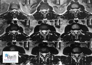
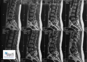
The patient is a young man with severe back pain, walking disorders and pain in the legs, especially the left leg. MRI images diagnosed problems in the fourth and fifth vertebrae. That’s why patient suffer from severe back pain. In dynamic photographs that imaging of patient taken in flexion, it became clear that the fourth and fifth vertebrae is quite unstable and wobbly. If we rely on conventional MRI, the patient’s condition was not detected correctly. This showed importance of accuracy and early detection of chronic back pain.
In this surgery Dr. Guive Sharifi (neurosurgeon) did laminectomy, vertebrae screws and Reduction dislocated vertebrae. The patient’s disk was drained. Between two vertebrae joined by use of intervertebral fusion, with own patient’s bone (autologous graft) .
Preoperative Imaging


video


